Interdisciplinary team wins grant to speed oral cancer testing
Published on Sep. 18, 2020
National Institutes of Health awards $420,000 to multidisciplinary team from Case Western Reserve University developing device for instantly detecting oral cancer
Mouth lesions are among the main early indicators of oral cancer, but determining whether a sore is actually malignant typically involves painful, costly biopsies.
Case Western Reserve researchers think they have a better idea—and the National Institutes of Health has given them $420,000 to advance it.
Highly collaborative in nature, the project teams researchers from the university’s schools of dental medicine, medicine and engineering.
Their concept combines imaging and algorithmic technologies to assess whether or not a lesion’s cells are malignant.
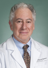
Aaron Weinberg, chair and professor in the Department of Biological Sciences in the School of Dental Medicine and professor of otolaryngology and pathology in the School of Medicine
“The idea is to quickly, easily, non-invasively and cost-effectively determine if a suspicious lesion is cancerous, or in other cases, to screen individuals on a regular basis to see if their lesion has progressed to cancer,” said dental faculty member Aaron Weinberg, the project’s principal investigator.
On average, oral cancer claims the life of someone in America every hour, according to the Oral Cancer Foundation.
Early detection is the key to survival, said Weinberg, chair and professor in the dental school’s Department of Biological Sciences. Their approach not only aims to reduce the need for biopsies, but also provide a much-needed diagnostic alternative in places where pathology services are few and difficult to access.
How it works
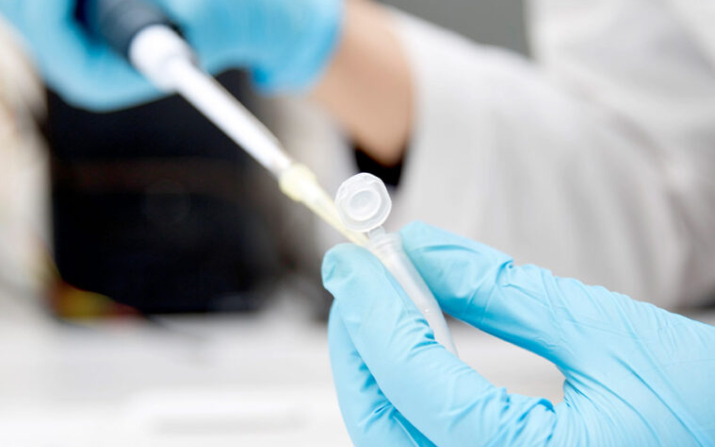
Researchers envision a hand-held device that can determine the ratios of two key proteins, where one goes up in cancer while the other one does not. By collecting cells swabbed from suspicious oral lesions, and using fluorescent antibodies that bind specifically to one or the other protein within the cells, the device collects images of fields of cells and an app converts the fluorescence intensity of each protein into numbers and calculates the ratio.
The numerical value above a given threshold indicates cancer.
This procedure has already proven to be accurate and effective in laboratory-based techniques, Weinberg said—and the intention is to convert the lab procedure into a device that will cut the time in obtaining results from two days to about 30 minutes.
“This will address major unmet needs in early oral cancer detection worldwide, especially in resource poor settings where pathology review is lacking and/or unreliable,” he said.
Weinberg is joined in the research by Umut Gurkan, the Warren E. Rupp Associate Professor at the Case School of Engineering; Anant Madabhushi, the F. Alex Nason Professor II of Biomedical Engineering and director of the Center for Computational Imaging and Personalized Diagnostics; and Rod Rezaee, associate professor in the Department of Otolaryngology at the School of Medicine and University Hospitals.
Director, Center for Computational Imaging and Personalized Diagnostics
Develops and translates computational imaging, AI and machine learning approaches for precision-medicine diagnosis, prognosis, treatment response and prediction