Histostitcher
Overview
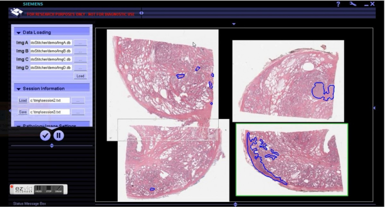
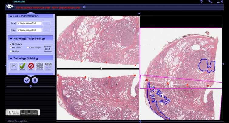
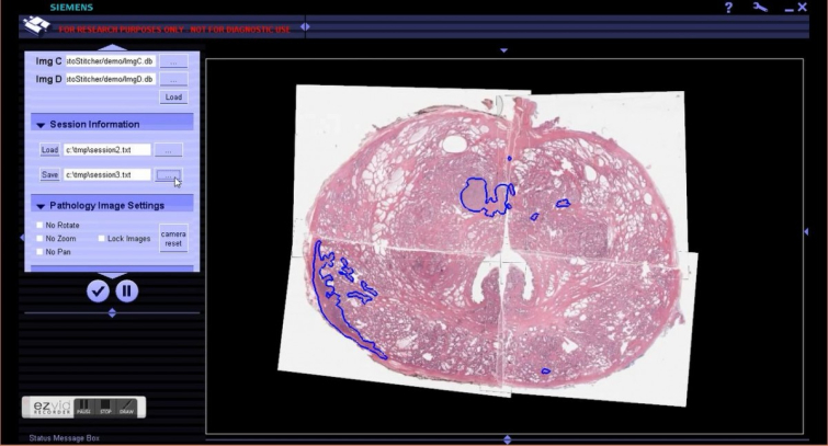
HistoStitcher is a free software package developed for reconstruction of whole mount histology sections (WHMS) from digitized tissue fragments. HistoStitcher allows users to select common fiducials on the edges of adjacent histology fragments and automatically determines the optimal transformations required to combine the images such that the selected points are maximally aligned. HistoStitcher also offers real time rendering of digital pathology images using multi-resolution pyramid to facilitate selection of fiducials, annotations of regions of interest and evaluation of the final stitch. This software is intended for research purposes only, not for diagnostic or clinical use.
Features:
(1) Data conversion tool to convert images to tile-based multi-resolution JPEG-encoded image format (db)
(2) Stitching via fiducial selection
(3) Manual image alignment without fiducial selection
(4) Importing, exporting, viewing, generating and stitching annotations corresponding to histologic fragments of interest
(5) Simultaneous viewing up to four high resolution, very large histologic images
Download
Manual
Installation
Requirements:
OpenGL 3.0 or later
Windows 7 64bit OS
Matlab runtime compiler 7.14 - included with Histostitcher installation
AMD SDK v 2.7- included with Histostitcher installation
*Windows Virtual Machines on Apple MacBooks are currently not supported*
Support and Contact
For questions or comment, please send here
Related Publications
Toth, Robert J., et al. "Histostitcher™: An informatics software platform for reconstructing whole-mount prostate histology using the extensible imaging platform framework." Journal of pathology informatics 5 (2014).
Toth, Robert, et al. "Incorporating the whole-mount prostate histology reconstruction program Histostitcher into the extensible imaging platform (XIP) framework." SPIE Medical Imaging. International Society for Optics and Photonics, 2012.
Chappelow, Jonathan, et al. "HistoStitcherTM: An interactive program for accurate and rapid reconstruction of digitized whole histological sections from tissue fragments." Computerized Medical Imaging and Graphics35.7 (2011): 557-567.
Funding Institutions
R01 Academic-Industrial grant (NIH R01CA136535-01)
Collaborators
University of Pennsylvania
Siemens Corporate Research
AutoStitcher
Overview
AutoStitcher is a free software program developed for reconstruction of whole mount histology sections (WHMS) from digitized tissue fragments. AutoStitcher takes as input a set of 4 digitized histology quadrants. This software is intended for research purposes only, not for diagnostic or clinical use.
For an example and instructions explaining how to run AutoStitcher, refer to documentation in the files AutoStitcher_example_script.m, and AutoStitcher.m.
Download
Matlab code available here
Related Publications
Penzias, G. et al. AutoStitcher: An Automated Program for Efficient and Robust Reconstruction of Digitized Whole Histological Sections from Tissue Fragments. Scientific Reports 6, 29906 (2016).
The publication is freely available for download at: http://www.nature.com/articles/srep29906
doi:10.1038/srep29906
Histoview
Overview
HistoView is a graphical user interface for pathology image analysis and visualization. It supports whole-slide images, provides a variety of automatic segmentation algorithms based on CCIPD’s literature and technical description of existing medical image analysis algorithms, and visualization of the segmentation result.
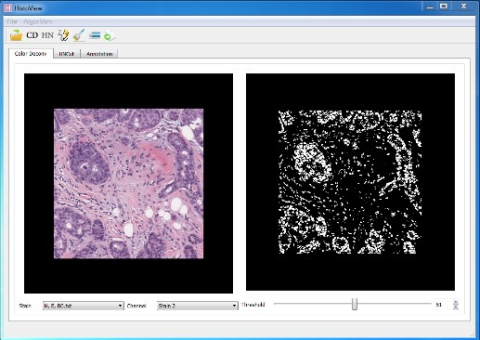
In HistoView v1.0, color deconvolution algorithm is embedded, which separates the image into different channels, corresponding to the actual colors of the stain used. User can change a threshold from 0 – 100 to binarize the channel image to measure specific tissues or structures.
Demo: https://www.youtube.com/watch?v=4XbbvZXNnLQ
Download
Histoview_download (Includes User guide and sample stains)
For questions or comment, please send here