Researchers aim artificial intelligence at rising oral cancers with $3.3 million grant from National Cancer Institute
Published on Jan. 5, 2021
Case Western Reserve and partners local and global team up to better predict which patients with oral squamous cell carcinomas will need radiation or chemotherapy after surgery
Researchers at Case Western Reserve University and partners in the United States and India are applying the investigative and predictive capabilities of artificial intelligence (AI) to help physicians customize treatments for patients with oral squamous cell carcinomas.
Research shows that oral squamous cell carcinomas cancer is already the eighth-most common type worldwide and numbers are steadily increasing in the United States, India and other parts of Asia.
The CCIPD has become a global leader in AI-driven precision medicine research. Madabhushi and his research team at the CCIPD hold more than 60 patents, many tied to their work in various cancers.
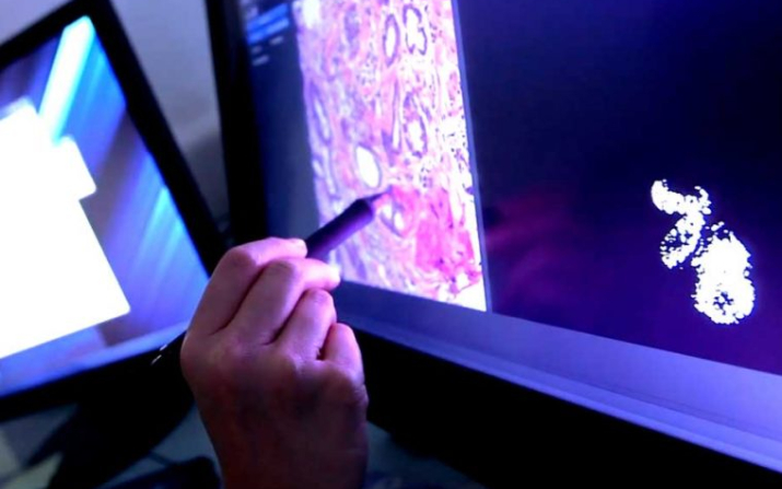
In this work, researchers will use advanced computer vision and machine learning techniques to identify cancer and immune cells on digitized images of oral squamous cell carcinoma tissue slides and then recognize spatial patterns among those cells.
This technology allows computerized vision to recognize patterns and quantify features that simply are beyond the human visual system but are powerful indicators of tumor biology. These algorithms will help oncologists and pathologists to then better determine which cancers are more versus less aggressive.
This, in turn, will then enable them to identify which patients with early-stage disease could safely receive surgery alone, versus who might need postoperative radiation. In addition, it could help identify which patients with advanced stage disease might need chemotherapy with radiation after initial treatment versus who may be adequately treated with radiation alone.
“We have known for a long time that pathologic features of oral cavity squamous cell carcinomas correlate with tumor behavior and prognosis, but human visual systems cannot extract these features consistently or quantitatively,” Lewis said. “AI now allows us to do just that, and we are hopeful that the extracted information can be turned into clinically available algorithms that drive better patient care decisions.”
Madabhushi and Lewis will work with a number of partners—Cleveland Clinic and University Hospitals in Cleveland, the San Francisco VA Health System, and Tata Memorial Centre in Mumbai, India—in a national and global endeavor to improve oral cavity squamous cell carcinoma patient care with advanced technology and data sharing.
The clinical partners will provide glass slides to be digitized or will directly provide digitally scanned whole slide images, which will be used to train the AI algorithms for predicting outcomes as well as treatment benefit.
The team will also have access to unique datasets from completed prospective, randomized, clinical trials of oral squamous cell carcinoma patients at the Tata Memorial Center as well as from the cancer clinical cooperative group NRG Oncology. The datasets that will allow for validation of the AI tools.
Seeking precise, personal predictions
Currently, physicians place oral carcinoma patients into one of three categories: those who require just surgery; those who should have surgery plus radiation therapy; or those who will need surgery, followed by radiation and chemotherapy.
“That’s the gold standard right now: a system that puts patients in those very broad categories,” Madabhushi said. “For clinicians and pathologists, this is limiting because it relies on a limited number of parameters. But our machines are looking at the appearance of cells, their spatial architecture and interplay between different cell types, to parse out those patients who should actually be in another category.”
For example, Madabhushi said, their AI research has already shown that there is a subset of early-stage patients now placed in the first category—surgery alone—who are actually at a much higher risk and would do poorly with surgery alone.
“Instead, they should be offered radiation therapy as well, but under the current parameters, that is not called for,” Madabhushi said.
The group also will look at anticipated differences in the appearance of oral cancer among patients of different races, a fast-developing aspect of Madabhushi’s AI-based investigations.
Oral carcinomas include cancers of the mouth, tongue, gums, and lips. According to the National Institutes of Health (NIH), these cancers can develop on the mobile tongue, the tissue lining the gums and hard palate, and on the underside of the tongue and floor of the mouth,
Oral carcinoma accounts for roughly 3% of all cancers diagnosed annually in the United States, with nearly 400,000 new cases being diagnosed annually worldwide.
Oral carcinoma most often occurs in people over age 40 and affects more than twice as many men as women. Most oral cancers are related to tobacco use, alcohol use, or both. Infection by the human papillomavirus (HPV), which is very common in oropharyngeal carcinomas, is a less common cause of oral carcinomas.
Other members of the research team include: Shlomo Koyfman, David Adelstein, and Deborah Chute at the Taussig Cancer Center, Cleveland Clinic; Ted Teknos, president of Seidman Cancer Center, University Hospitals; Stephen Connelly, San Francisco VA Health System; and Sarbani Ghosh-Laskar and Swapnil Rane, Tata Cancer Center.
Director, Center for Computational Imaging and Personalized Diagnostics
Develops and translates computational imaging, AI and machine learning approaches for precision-medicine diagnosis, prognosis, treatment response and prediction